

RESEARCH NOTE
Anne Voss, Ronald Stone, and John Scott Foott*
U.S. Fish and Wildlife Service, Pacific Southwest Region, 24411 Coleman Hatchery Road, Anderson, CA 96007 https://orcid.org/0000-0003-0892-6425 (JSF)
Published 8 October 2026 • doi.org/10.51492/cfwj.112.12
Key words: Ceratonova shasta, Chinook salmon, fish disease, Klamath River, Oncorhynchus tshawytscha, Trinity River
| Citation: Voss, A., R. Stone, and J. S. Foott. 2026. Ceratonova shasta infection of Trinity River Chinook salmon smolts: another factor in low recruitment. California Fish and Wildlife Journal 112:e12. |
| Editor: John Kelly, Fisheries Branch |
| Submitted: 8 December 2025; Accepted: 23 March 2026 |
| Copyright: ©2026, Voss et al. This is an open access article and is considered public domain. Users have the right to read, download, copy, distribute, print, search, or link to the full texts of articles in this journal, crawl them for indexing, pass them as data to software, or use them for any other lawful purpose, provided the authors and the California Department of Fish and Wildlife are acknowledged. |
| Funding: Partial funding for this work came from the Bureau of Reclamation Trinity Restoration Program and Klamath basin area office interagency agreements, and the U.S. Fish and Wildlife Service National Wild Fish Survey. |
| Competing Interests: The authors have not declared any competing interests. |
The Trinity River, a 266-km tributary of the Klamath River in northern California, is regulated by the Trinity and Lewiston dams, which were constructed in the 1960s as part of the federal Central Valley Project. These dams permanently blocked upstream migration routes for salmon and steelhead trout, significantly impacting native fish populations. To offset these impacts and support the continued presence of anadromous fish in the basin, the California Department of Fish and Wildlife (CDFW) operates the Trinity River Hatchery (TRH). The hatchery produces spring-run and fall-run Chinook salmon (Oncorhynchus tshawytscha), Coho Salmon (O. kisutch), and steelhead (O. mykiss) to help sustain fish populations, support tribal and recreational fisheries, and fulfill mitigation obligations associated with dam construction. In the past decade, adult Trinity River Chinook salmon returns have declined significantly, resulting in harvest restrictions beginning in 2022 (Sullivan and Hileman 2019). The decline of California’s Chinook salmon populations is the result of multiple, interacting stressors. Key contributing factors include historical overfishing, predation by non-native species, degraded water quality, physical barriers and habitat degradation from legacy mining activities, and the loss of spawning and rearing habitat due to dams and water diversions. Additionally, altered flow regimes, ecological interactions between hatchery-origin and natural-origin fish, and disease have further compounded population declines. (Hankin 1985; Yoshiyama et al. 2001; Williams et al. 2011; Sullivan and Hileman 2019; Sturrock et al. 2019; Lehman et al. 2020). Infectious disease has been recognized as an important factor for Pacific Northwest salmonid population survival (Zuray et al. 2012; Miller et al. 2014; Furey et al. 2021; Foott et al. 2023).
Ceratonova shasta (formerly Ceratomyxa shasta; Atkinson et al. 2014) is a freshwater myxozoan parasite of Pacific Northwest salmonids and has been reported as a major disease agent in the Klamath River (Foott et al. 2004; Stocking et al. 2006; Fujiwara et al. 2011; Hallett et al. 2012; Atkinson et al. 2014). Ceratonova shasta has a complex life cycle, involving both an invertebrate annelid host (Manayunkia occidentalis) and a vertebrate salmonid host (Bartholomew et al. 1997; Atkinson et al. 2020). Infected annelids release actinospores into the aquatic environment where the spores attach to the salmon gill epithelium, invade the circulatory system, replicate, and subsequently migrate to the intestinal tract for further multiplication and sporogony (Bjork and Bartholomew 2010). Infected fish may develop enteritis (inflammation of the intestinal tract) with varying severity and associated anemia based upon factors such as parasite genotype and density, innate host resistance, and water temperature (Udey et al. 1975; Foott et al. 2004; Bjork and Bartholomew 2009; Ray et al. 2010; Hallett et al. 2012; Bartholomew et al. 2022). The myxospore stage develops within the salmon and is typically released post-mortem (Foott et al. 2016). If ingested by the filter-feeding annelid, the parasite completes its life cycle by invading the annelid’s gut epithelium (Meaders and Hendrickson 2009). Another myxozoan, Parvicapsula minibicornis, infects the same annelid and salmonid hosts as C. shasta and is frequently observed as a co-infection in salmon (Bartholomew et al. 2006). Parvicapsula minibicornis exhibits a tropism for the kidney nephron and can induce glomerulonephritis in the salmon host (Foott et al. 2004). While glomerulonephritis is an additive stressor, it is our hypothesis that the hemorrhagic enteronecrosis associated with C. shasta infection is the primary driver of pathogen related mortality for juvenile salmon, exceeding the impact of P. minibicornis. A general microbiological and parasite focused survey was conducted on natural and hatchery juvenile Chinook salmon within the Trinity River (1991–2021). Beginning in 2009, we used a molecular diagnostic technique to focus on C. shasta infection of salmon in the Trinity River and coded wire tagged TRH smolts in the Klamath estuary.
The Trinity River Hatchery is located at rkm 180, situated at the base of Lewiston Dam which marks the upstream limit of anadromous fish migration. From the hatchery, the mainstem Trinity River flows 180 km downstream to the confluence with the Klamath River (rkm 0). Out migrating juvenile salmon move past the Pear Tree trap site (rkm 118) and the Willow Creek trap site (rkm 34) before entering the Klamath River. The migration continues through the lower Klamath River, which is divided into the K1 reach (rkm 70.8 to 25.7) and the K0 reach (rkm 25.7 to the Pacific Ocean).
We collected fish from four locations. Sampling occurred at TRH raceways, rotary screw traps operated at Pear Tree (2021, PCT) and Willow Creek (1991–2021, WCT), and by beach seine within the K0 estuary reach of the Klamath River (2009–2025; Fig. 1). In 1998 and 2002, sampling focused on moribund fish (diagnostic approach) at the above sample sites.
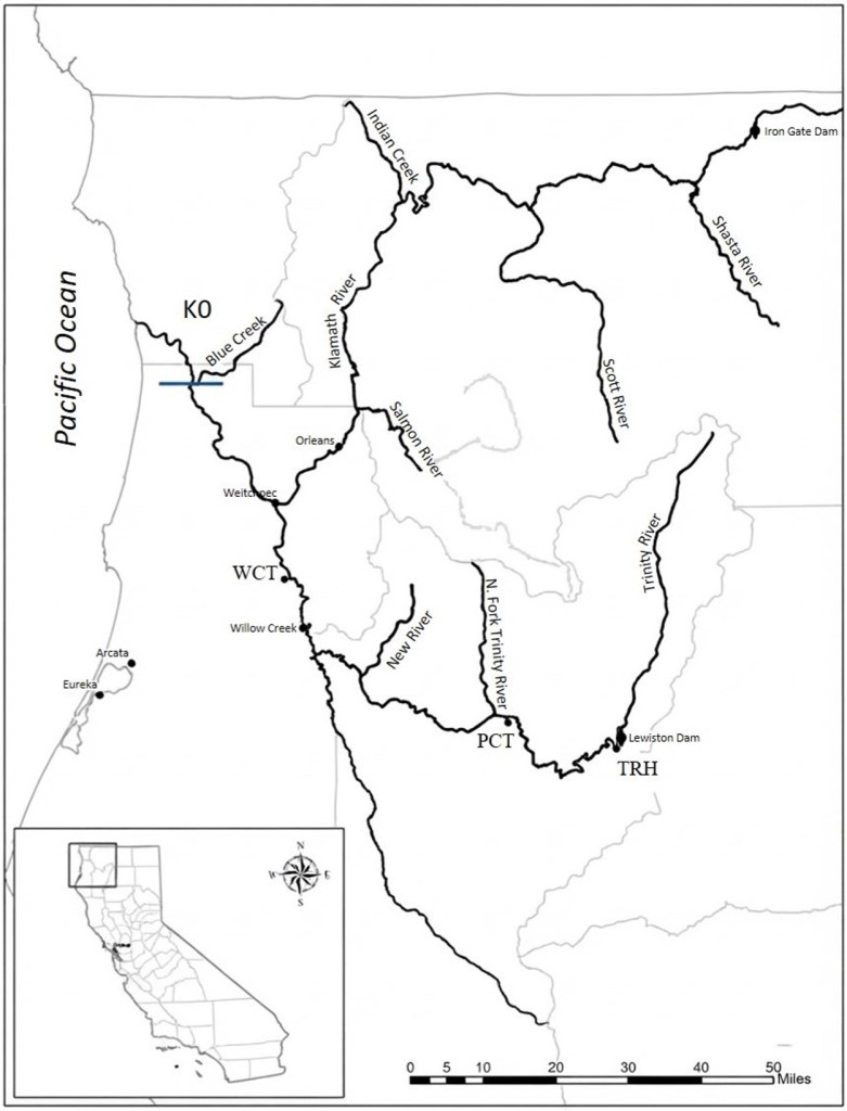
We defined natural fish as unmarked salmon (intact adipose fin) captured prior to the annual TRH release. The Trinity River Hatchery conducted volitional releases of Chinook salmon in late May–June (spring smolt) and in October (yearlings, age 1+). Both release groups include a 25% coded wire tag (CWT) mark. We identified hatchery origin by dissecting coded wire tags from adipose fin-clipped fish and comparing the tag codes to release records provided by TRH.
In 1991, 1992, and 2021, we aseptically collected the anterior kidney for cultured bacteria (Brain Heart Infusion agar at 22º C incubation) and virus (inoculation onto EPC and CHSE214 cell lines at 15⁰ C; USFWS and AFS-FHS 2014). We classified gram-negative, motile, no diffusible pigment, oxidase positive bacteria as motile Aeromonad bacteria (Aeromonas sp./ Pseudomonas sp. (AP) (Roberts 1989). Gills from a subset of affected fish were examined by either gram-stained imprint or cultured on Tryptase Yeast Extracts Salts agar for filamentous gram-negative rods and were presumptively identified as Flavobacterium columnare. Renibacterium salmoninarum infection of the kidney was assayed by two methods, direct fluorescent antibody method (1991–1993, and 2021) or ELISA (1992–1996; USFWS and AFS-FHS 2014). For the R. salmoninarum ELISA, we diluted kidney tissue 4x (w/v) in PBS-0.05% tween20, homogenized, boiled for 15 minutes, centrifuged, and assessed the supernatant for antigen using commercial polyclonal antibody (Kirkegaard and Perry Laboratories, Inc. Gaithersburg, MD, USA; Pascho and Mulcahy 1987). A positive threshold was set conservatively at an optical density of 0.18. This value was the mean OD405 of the lowest antigen standard (1:5000) and represented a 2.0 signal to noise ratio (mean OD405 of the lowest antigen standard (1:5000)/mean of the negative kidney control) (Pascho and Mulcahy 1987).
In 1992–1996, we determined metacercaria infection of the kidney by pressing kidney tissue between two microscopic slides and viewing the preparation with a binocular dissection microscope at 50–70X magnification. In 1994, we recorded the weight of the kidney tissue (0.001 g) and the number of metacercaria to obtain metacercaria per gram of kidney data.
We performed histology on salmon from 1991–2021, which was the sole method for detection of C. shasta and P. minibicornis diagnosis until 2009. Kidney, gill, and gastrointestinal tract were placed in Davidson’s fixative for 48 hours, processed for 5μm paraffin sections, and stained with hematoxylin and eosin (Humason 1979). In 1991, blood smears were collected, fixed in absolute methanol, stained with pinacyanol chloride and examined microscopically for erythrocytic viral inclusions (Leek 1987; Foott and Walker 1992).
Beginning in 2009, we collected intestine (anus to the junction of the small intestine with the pyloric ceca) with separate sterile tools and extracted DNA with Applied Biosystems MagMax Express-96 Magnetic Particle Processor or ThermoFisher Kingfisher Flex instrument. A modified C. shasta QPCR assay targeting the 18S ribosomal DNA sequence was used to assay the DNA extracted intestinal samples (Hallett and Bartholomew 2006). In 2009–2012, we also sampled DNA extracted from the kidney for a P. minibicornis QPCR assay (True et al. 2009). All reactions (30 µL) were performed in a 96-well optical reaction plate using 0.9 µM of both primers, 0.25 µM of probe, 15 µL of TaqMan Universal PCR Master Mix, and 5µL of DNA template. The DNA template volume was increased from one µL in the Hallett and Bartholomew (2006) paper to reduce pipetting error of small volumes. We based primer and probe concentrations on default settings for the Applied Biosystems readers and tested samples in a single well without an internal positive control. Reactions were assayed using a 7300 Sequence Detection System (Applied Biosystems) from 2006–2021 and the QuantStudio 6 real-time PCR system (ThermoFisher) since 2022 (Voss et al. 2023). Cycling conditions for all assays were the following: 2 min. at 50°C and 10 min. at 95°C, followed by 40 cycles of 95°C for 15 seconds and 60°C for one min. We examined amplification plots (cycle vs. fluorescent signal) to verify appropriate baseline crossing, a steep exponential amplification phase, and a clear plateau.
We reviewed multicomponent plots to confirm increasing reporter dye fluorescence with stable passive reference dye signal. Samples displaying abnormal amplification curves, suggestive of inhibition, were diluted and reanalyzed. Each assay plate included a standard curve with three concentrations of reference standards (two replicates each) at known DNA copy number and two negative control wells. We obtained the C. shasta reference standard curve using synthesized DNA (gBlock Gene Fragments, Integrated DNA Technology, Coralville, IA, USA) containing the 18S ribosomal DNA target sequence (Voss et al. 2022). The P. minibicornis standard was a plasmid DNA control (pPM-q18S, 3943 bp) with a molecular weight (MW) of 2,395,136 g mol–1. Using this MW, we estimated that 1 µg of plasmid DNA contains 2.5 x 10¹¹ gene copies. Using QPCR analysis software, we calculated the cycle threshold (CT) values for each standard concentration (SDS software 7300 SDS v 1.4, QuantStudio 6 v. 1.7.2, Applied Biosystems). Criteria for a positive test result required samples to produce a change in normalized fluorescent signal (Δ Rn) greater than or equal to 100,000 fluorescent units and ≥ 5 copies (≥ 40 plasmid copies for P. minibicornis). We evaluated each assay for expected CT values of the reference standards and assay efficiency. At the end of the season, any plates with more than a 3% decrease in assay efficiency from the mean were retested and reevaluated. Low copy numbers are not reliable given the limitations of the Poisson distribution (Applied Biosystems 2016). Ceratonova shasta DNA copy (log) was obtained for each sample based on a known quantity from the standard curve. We defined three logs or greater of C. shasta DNA as the threshold for a diseased state (infections likely to lead to mortality under spring-summer temperatures). This threshold, referred to as CS2, was based on a sentinel exposure study conducted in 2008 (True et al. 2012) and QPCR–histology comparisons (Voss et al. 2022). A “CS1” rating meant the intestinal sample was positive but less than 3.0 logs of C. shasta DNA. Prevalence of infection (POI) is the percentage of positive samples among total samples. We do not have similar data for P. minibicornis copy number and level of glomerulonephritis (True et al. 2009).
Most fall-run Klamath Basin Chinook salmon spawning adults are three years old (Klamath River Technical Team 2021). Using CDFW grilse (two years old) and adult returns data to TRH, (CDFW 2025, Megatable documents), we related TRH smolt CS-POI to adult returns two years later (assumes the majority are 3-year-old salmon). We examined the relationship between infection prevalence of TRH smolts captured in the estuary and subsequent adult return by the Pearson product-moment correlation coefficient (R) test.
A total of 2,076 juvenile Chinook salmon were sampled within the Trinity River over eleven survey years (1991–1994, 1996, 1998, 2001, 2002, 2011, 2012, and 2021), including 869 naturals (collected May and June prior to hatchery releases), 457 TRH spring and 426 fall releases (CWT), and 324 TRH preliberation fish. Of those, we analyzed 1,862 salmon by QPCR for C. shasta (2009–2025) and tested 563 fish for P. minibicornis (2009–2012).
Systemic bacterial infections were infrequent. Motile aeromonads were the most common, with prevalence ranging 6–26% (Table 1). Eroded gills, containing gram-negative filamentous bacteria, were observed in otherwise normal appearing smolts at WCT during diagnostic evaluations when river temperatures were ≥ 21⁰C (57% in 1998, 2% POI in 2001). We observed yellow-orange rhizoid colonies of gram-negative filamentous bacteria on Tryptase Yeast Extracts Salts agar cultures from a subset of gill samples and presumptively identified as Flavobacterium columnare. Infectious Hematopoietic Necrosis Virus was isolated from TRH salmon in the May 1991 preliberation sample, and the hatchery had experienced an epizootic due to the virus that season. No virus was isolated from any 1991 down-river outmigrants or any other salmon sampled through 2021.
Table 1. Prevalence of infection for parasites, bacteria, and virus in natural and Trinity River Hatchery (TRH) juvenile fall-run and spring-run Chinook salmon sampled in-river (adipose fin clip with TRH coded wire tag, CWT) or at the hatchery prior to release (Prelib.) in 1991–1994, 1996, 2011–2012, and 2021. TRH spring release (rel.) were age 0 smolts and fall release were yearlings. Assays included both direct fluorescent antibody (DFAT) and enzyme-linked immunosorbent assay (ELISA) for Renibacterium salmoninarum, microscopy of kidney squash wet mount preparations (metacercaria) and stained blood smears (erythrocytic (RBC) inclusions), kidney inoculum onto Brain Heart Infusion agar (cultured bacteria) and tissue culture (virus), and microscopic evaluation of histology sections of various tissues for presumptive parasite identification. ND = not done
| Sample Location | Natural | TRH CWT Spring Release | TRH CWT Fall Release | TRH Prelib. |
| Kidney R. salmoninarum DFAT | 1/333 (0.3%) | 3/166 (2%) | ND | 0/42 (0%) |
| Kidney R. salmoninarum ELISA | 9/238 (4%) | 5/185 (3%) | 22/411 (5%) | 10/219 (5%) |
| Kidney squash metacercaria | 250/341 (73%) | 148/195 (76%) | 263/331 (79%) | 3/63 (5%) |
| Cultured bacteria aeromonids | 8/130 (6%) | 18/70 (26%) | ND | 8/60 (13%) |
| Virus | 0/152 (0%) | 0/15 (0%) | ND | 3/79 (4%) |
| RBC inclusions | 0/92 (0%) | 0/40 | ND | 0/59 (0%) |
| Histology (intestine) C. shasta | 0/279 (0%) | 0/47 (0%) | 0/15 (0%) | 0/22 (0%) |
| Histology (kidney) Metacercaria | 21/195 (11%) | 29/67 (43%) | 0/14 (0%) | 0/24 (0%) |
| Histology (kidney) P. minibicornis | 0/195 (0%) | 0/67 (0%) | 0/14 (0%) | 0/24 (0%) |
| Histology (kidney) Chloromyxum sp.* | 14/195 (7%) | 0/67 (0%) | 3/14 (21%) | 0/24 (0%) |
| Histology (gill) Glochidia | 25/264 (9%) | 13/96 (14%) | 0/15 (0%) | 0/26 (0%) |
| Histology (gill) Metacercaria | 48/264 (18%) | 15/96 (16%) | 0/15 (0%) | 0/26 (0%) |
* Presumptive identification based on morphology and tissue location
Metacercaria infection of the kidney was the most common observation, with prevalence ≥ 73% in Trinity River outmigrant salmon (Table 1). This trematode was only observed at TRH in 1993. Metacercaria were presumptively identified as Nanophyetus salmonicola, based on the morphology of the metacercaria with its prominent excretory vesicle (Fig. 2), the presence of the intermediate host Juga sp. snail, and the trematode’s published range (Millemann and Knapp 1970; Pennak 1989). We did not perform molecular confirmation as this diagnostic technique was unavailable at the Fish Health Center when kidney squash counts were performed. In the spring of 1994, both natural and hatchery salmon had metacercaria prevalence of > 75% infection with the mean metacercaria/g kidney of 199 or greater (Table 2). Within one week of release into the Trinity River, hatchery salmon had a 79% POI. Given the size of these salmon (< 85 mm), the maximum count per gram of 5,464 is quite striking. Despite high parasite loads, none of the sampled fish showed signs of morbidity or external signs of infection. Previous studies indicate pathogenesis varies by fish species-specific resistance, rate of accumulation, and water temperature (Baldwin et al. 1967; Jacobson et al. 2008). TRH yearling fall-run Chinook salmon exposed for 27 days to infected Juga sp. snails developed infections ranging from 32 to 10,220 metacercaria per gram of kidney tissue, with over 50% of the infected fish having > 5,000 metacercaria/g (Foott et al. 1997). No mortality occurred to these infected fish; however, they showed an increase in immunoglobulin and a decrease in saltwater osmoregulatory capacity. It is unclear what the disease prognosis is for these infected Trinity River juvenile salmon. Encysted metacercaria were also observed in gill and kidney sections from both natural and TRH CWT salmon during the spring (Table 1).
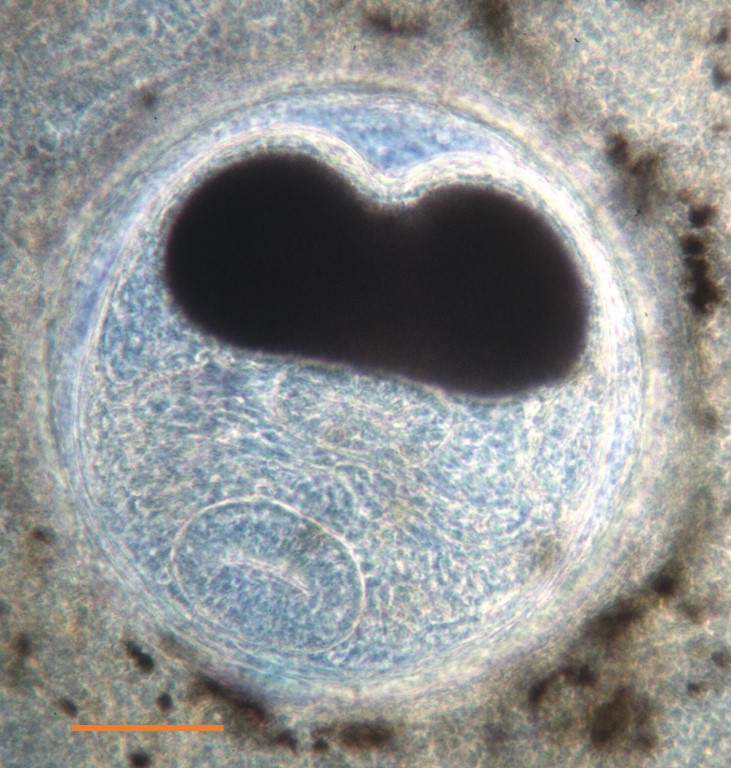
Table 2. Metacercaria kidney infection of juvenile natural and hatchery (TRH-CWT-week post release) captured at the Willow Creek trap in June 1994. Prevalence of infection (POI = number positive/total sample), mean count per g kidney ± SEM, and maximum count/g kidney.
| Capture Location, Date | POI | Mean | Max |
| Natural, 30 May–8 June | 79/105 (75%) | 296 ± 37 | 2076 |
| TRH CWT-1, 16 June | 22/28 (79%) | 199 ± 32 | 565 |
| TRH CWT-2, 21 June | 35/35 (100%) | 520 ± 158 | 5464 |
| TRH CWT-3, 28 June | 57/65 (88%) | 302 ± 44 | 1589 |
No erythrocytic inclusions were seen in blood smears from 191 Chinook salmon smolts examined in 1991. Freshwater mussel glochidia were observed in natural and TRH CWT salmon gills during the spring (Fig. 3). The western pearl shell mussel (Margaritifera falcata) is reported to be the only mussel in the Trinity River (Felbeck and Hauser 2020). Presumptive Chloromyxum sp. (myxozoan parasite) was observed in the Bowman’s capsule of a low prevalence of salmon kidneys (Fig. 4). Histological analysis did not detect C. shasta (363 intestine sections) or P. minibicornis (300 kidney sections) in TRH pre-release, out migrating TRH CWT, or natural Chinook salmon (Table 1).


Quantitative PCR, a more sensitive assay than histology, detected both C. shasta and P. minibicornis in coded-wire tagged TRH Chinook salmon collected at the hatchery (2009) and in-river (primarily in 2012; Table 3). In spring of 2021, neither C. shasta nor P. minibicornis was detected in 113 natural juveniles collected at Pear Tree and Willow Creek traps and assayed by QPCR (54 fish) or histology (59 fish). DNA copy numbers were less than 3 logs in the C. shasta positive fish indicating asymptomatic infections. While limited to four years (2010–2012, 2021) , these data indicate that while both C. shasta and P. minibicornis are present in the Trinity River; they are at low abundance in comparison to the Klamath River (Bartholomew et al. 2007; Hallett et al. 2012; Robinson et al. 2020).
Table 3. Prevalence of infection for Ceratonova shasta (intestine) and Parvicapsula minibicornis (kidney) assayed by QPCR from Trinity River hatchery fall and spring-run Chinook salmon smolts sampled at the hatchery prior to their spring release (TRH) or code-wire salmon collected in the Trinity R. post-release (in-river CWT).
| Release | Year | C. shasta | P. minibicornis |
| TRH | 2009 | 9/48 (19%) | 12/48 (25%) |
| TRH | 2010 | 0/70 (0%) | 0/70 (0%) |
| TRH | 2011 | 0/62 (0%) | 0/62 (0%) |
| TRH | 2012 | 0/30 (0%) | 0/30 (0%) |
| In-river CWT | 2010 | 0/216 (0%) | 2/216 (1%) |
| In-river CWT | 2011 | 0/117 (0%) | Not tested |
| In-river CWT | 2012 | 9/137 (7%) | 52/137 (53%) |
Prevalence of C. shasta infection (CS-POI), in fall and spring-run TRH CWT smolts captured within the estuary ranged from 3–85% over the 14-year survey period (Table 4). Estuary TRH CWT smolts were likely exposed to the Klamath River for one week or more (Klamath River Basin Juvenile Salmonid Investigations 2000). The infectious “dose” that is likely to result in clinical disease has been reported as a C. shasta spore/L concentration ≥ 10 (Hallett et al. 2012). Water samples collected in June 2025 in the Klamath River below the confluence with the Trinity River (mouth of Tully Creek, rkm 62.55) contained 10–20 spores/L, corroborating C. shasta infections observed in TRH smolts (OSU 2025). Most infected smolts were classified as CS1 (infected but not likely in a disease state at the time of collection). Over eight survey years (2013–2015, 2021–2025), CS-POI exceeded 50%, and three years (2013, 2021–2023), had ≥ 26% of smolts classified as CS2 (“diseased”; Fig. 4). Ceratomyxosis is a progressive disease and continues in saltwater (Ching and Munday 1984; Bartholomew et al. 1989). The disease prognosis for the majority of infected smolts was likely enteronecrosis and anemia (Foott et al. 2004; Bjork and Bartholomew 2010).
Table 4. Prevalence of infection for Ceratonova shasta (CS-POI = CS1+CS2/total sample) assayed by QPCR from Trinity River hatchery code-wire tagged fall and spring-run Chinook salmon smolts sampled in the Klamath estuary during the spring of 2009–2015, 2018–2019, and 2021–2025. Severity of infection reported as number of fish rated at CS1 (infected but less than 3 copies of parasite DNA) and CS2 (≥ 3 copies of parasite DNA).
| Year | CS1 | CS2 | CS-POI |
| 2009 | 5 | 2 | 7/81 (9%) |
| 2010 | 1 | 0 | 1/36 (3%) |
| 2011 | 2 | 0 | 2/21 (10%) |
| 2012 | 17 | 4 | 21/49 (43%) |
| 2013 | 1 | 32 | 33/55 (60%) |
| 2014 | 64 | 21 | 85/115 (74%) |
| 2015 | 6 | 1 | 7/12 (58%) |
| 2018 | 9 | 1 | 10/103 (10%) |
| 2019 | 36 | 8 | 44/118 (37%) |
| 2021 | 86 | 26 | 112/140 (80%) |
| 2022 | 41 | 26 | 67/79 (85%) |
| 2023 | 32 | 28 | 60/93 (65%) |
| 2024 | 90 | 19 | 109/161 (68%) |
| 2025 | 59 | 12 | 71/119 (60%) |
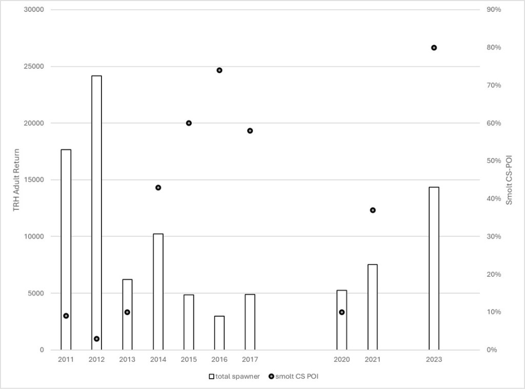
Over ten years, there was a negative relationship between smolt CS POI and adult return to TRH (R –0.404; Fig. 5). Three outlier years (2013, 2020, 2023) weakened this relationship. Removing these outliers increased R to –0.938. Potential explanations for two of these outliers could be closure of ocean harvest in 2022 (2023 returns) and poor outmigration conditions (2020 returns) due to the critically dry water year in the Trinity River (Trinity River Restoration Program 2025).
Ceratonova shasta has been reported as a significant disease affecting juvenile Chinook salmon in the Klamath River (Bartholomew et al. 2023), and juvenile Trinity River salmon must pass through the lower Klamath River during their smolt migration. Although QPCR results confirm that C. shasta is present in the Trinity River, data from four years indicated that infection levels were low relative to the Klamath River and predominantly asymptomatic. In contrast, smolts experience substantially higher exposure to C. shasta once they enter the lower Klamath River, where infectious actinospore concentrations are frequently at thresholds associated with a disease state. While multiple factors influence declines in Trinity River Chinook salmon including ocean harvest, elevated water temperature associated with low river flows, and insufficient upwelling resulting in low ocean productivity for smolts (Fisher and Pearcy 1988; Quinones et al. 2014), we believe ceratomyxosis may contribute to reduced Trinity River smolt survival rates during certain years. Given that out migrating juvenile salmon from the Trinity River must travel through the lower Klamath River, C. shasta should be included among potential factors when basin managers are evaluating drivers of adult returns. We suggest that monitoring for C. shasta in natural and coded wire tagged hatchery smolts within the lower Trinity River as well as tagged hatchery salmon in the Klamath estuary become a regular program.
Acknowledgments
We thank the fish biologists with the US Fish and Wildlife Service, Yurok and Hoopa tribes, and the California Department of Fish and Wildlife who assisted us in fish collection, and members of the Fish Health Laboratory (J. Williamson, K. True, K. Nichols, J. Jacobs, and S. Freund) for laboratory assistance. Partial funding for this work came from the Bureau of Reclamation Trinity Restoration Program and Klamath basin area office interagency agreements, and the U.S. Fish and Wildlife Service National Wild Fish Survey.
Data Availability
The data that supports the findings in this review are available from the corresponding author upon reasonable request.
Ethics Statement
Fish were euthanized following published guidelines (Use of Fishes in Research Committee 2014).
Literature Cited
- Applied Biosystems. 2016. Real-time PCR: Understanding Ct. Publication CO019879 0116. Thermo Fisher Scientific, Waltham, MA, USA. Available from: https://www.thermofisher.com/content/dam/LifeTech/Documents/PDFs/PG1503-PJ9169-CO019879-Re-brand-Real-Time-PCR-Understanding-Ct-Value-Americas-FHR.pdf (Accessed Sept 2018)
- Atkinson, S. D., J. S. Foott, and J. L. Bartholomew. 2014. Erection of Ceratonova n. gen. (Myxosporea: Ceratomyxidae) to encompass histozoic species C. gasterostea N. Sp. from three spine stickleback (Gasterosteus aculeatus) and C. shasta n. comb. from salmonid fishes. Journal of Parasitology 100(5):640–645.
- Atkinson, S. D., J. L. Bartholomew, and G. W. Rouse. 2020. The invertebrate host of salmonid fish parasites Ceratonova shasta and Parvicapsula minibicornis (Cnidaria) is a novel fabriciid annelid. Manayunkia occidentalis sp. nov. (Sabellida: Fabriciidae). Zootaxa 4751(2):310–320.
- Baldwin, N. L., R. E. Millemann, and S.E. Knapp. 1967. “Salmon poisoning” disease. III. Effects of experimental Nanophyetus salmincola infection on the fish host. Journal of Parasitology 53(3):556–564.
- Bartholomew, J. L., M. Whipple, D. Stevens, and J. L. Fryer. 1997. The life cycle of Ceratomyxa shasta, a myxosporean parasite of salmonids, requires a freshwater polychaete as an alternate host. Journal of Parasitology 83:859–868.
- Bartholomew, J. L., C. E. Smith, J. S. Rohovec and J. L Fryer. 1989. Characteristics of a host response to the myxosporean parasite, Ceratomyxa shasta (Noble), by histology, scanning electron microscopy and immunological techniques. Journal of Fish Diseases 12:509–522.
- Bartholomew, J. L., S. D. Atkinson, and S. L. Hallett. 2006. Involvement of Manayunkia speciosa (Annelida: Polychaeta: Sabellidae) in the life cycle of Parvicapsula minibicornis, a myxozoan parasite of Pacific Salmon. Journal of Parasitology 92:742–748.
- Bartholomew, J. L., S. D. Atkinson, S. L., Hallett, C. M. Zielinski, and J. S. Foott. 2007. Distribution and abundance of the salmonid parasite Parvicapsula minibicornis (Myxozoa) in the Klamath River basin (Oregon–California, USA). Diseases of Aquatic Organisms 78:137–146.
- Bartholomew, J. L., J. D. Alexander, S. L. Hallett, G. Alama-Bermejo, and S. D. Atkinson. 2022. Ceratonova shasta: a cnidarian parasite of annelids and salmonids. Parasitology 149(14):1862–1875.
- Bartholomew, J. L., J. D. Alexander, J. Alvarez, S. D. Atkinson, M. Belchik, S. J. Bjork, J. S. Foott, A. Gonyaw, M. E. Hereford, R. A. Holt, B. McCovey Jr., N. A. Som, T. Soto, A. Voss, T. H. Williams, T. G. Wise, and S. L. Hallett. 2023. Deconstructing dams and disease: predictions for salmon disease risk following Klamath River dam removals. Frontiers in Ecology and Evolution 11:1245967. https://doi.org/10.3389/fevo.2023.1245967
- Bjork, S. J., and J. L. Bartholomew. 2009. Effects of Ceratomyxa shasta dose on a susceptible strain of rainbow trout and comparatively resistant Chinook and coho salmon. Diseases of Aquatic Organisms 86:29–37.
- Bjork, S. J., and J. L. Bartholomew. 2010. Invasion of Ceratomyxa shasta (Myxozoa) and comparison of migration to the intestine between susceptible and resistant fish host. International Journal of Parasitology 40:1087–1095.
- California Department of Fish and Wildlife (CDFW). 2025. Chinook Salmon: Anadromous Assessment. Available from: https://wildlife.ca.gov/Conservation/Fishes/Chinook-Salmon/Anadromous-Assessment (Accessed: Nov 2025)
- Ching, H. L., and D. R. Munday. 1984. Susceptibility of six Fraser Chinook salmon stocks to Ceratomyxa shasta and the effects of salinity on ceratomyxosis. Canadian Journal of Zoology 62(6):1081–1083.
- Felbeck, F., and K. Hauser. 2020. Qualitative survey of western pearl shell mussel populations in the Trinity River. Bureau of Land Management, Redding Field Office, Redding, CA, USA. Available from: https://www.trrp.net/DataPort/doc.php?id=2573
- Fisher, J. P., and W. G Pearcy. 1988. Growth of juvenile coho salmon (Oncorhynchus kisutch) off Oregon and Washington, U.S.A., in years of differing coastal upwelling. Canadian Journal of Fisheries and Aquatic Sciences 45:1036–1044. https://doi.org/10.1139/f88‑127
- Foott, J. S., and R. L. Walker. 1992. Disease survey of Trinity River salmonid smolt populations. U.S. Fish and Wildlife Service, California–Nevada Fish Health Center, Anderson, CA, USA.
- Foott, J. S, D. Free, W. Talo, and J. D. Williamson. 1997. FY96 Investigational Report: Physiological Effects of Nanophyetus Metacercaria Infection in Chinook Salmon Smolts (Trinity River). U. S. Fish and Wildlife Service, California–Nevada Fish Health Center, Anderson, CA, USA.
- Foott, J. S., R. Harmon, and R. Stone. 2004. Effect of water temperature on non-specific immune function and ceratomyxosis in juvenile Chinook salmon and steelhead from the Klamath River. California Fish and Game 90:71–90.
- Foott, J. S., R. Stone, R. Fogerty, K. True, A. Bolick, J. L. Bartholomew, S. L. Hallett, G. R. Buckles, and J. D. Alexander. 2016. Production of Ceratonova shasta myxospores from salmon carcasses: carcass removal is not a viable management option. Journal of Aquatic Animal Health 28:75–84.
- Foott, J. S., J. Kindopp, K. Gordon, A. Imrie, and K. Hikey. 2023. Ceratonova shasta infection in lower Feather River Chinook juveniles and trends in water-borne spore stages. California Fish and Wildlife Journal 109:e9.
- Fujiwara, M., M. S. Mohr, A. Greenberg, J. S. Foott, and J. L. Bartholomew. 2011. Effects of ceratomyxosis on population dynamics on Klamath Fall-run Chinook Salmon. Transactions of the American Fisheries Society 140:1380–1391.
- Furey, N. B., A. L. Bass, K. M. Miller, S. Li, A. G. Lotto, S. J. Healy, S. M. Drenner, and S. G. Hinch. 2021. Infected juvenile salmon can experience increased predation during freshwater migration. Royal Society Open Science 8:201522.
- Hallett, S. L., and J. L. Bartholomew. 2006. Application of a real-time PCR assay to detect and quantify the myxozoan parasite Ceratomyxa shasta in water samples. Diseases of Aquatic Organisms 71:109–118.
- Hallett, S. L., R. A. Ray, C. N. Hurst, R. A. Holt, G. R. Buckles, S. D. Atkinson, and J. L. Bartholomew. 2012. Density of the waterborne parasite Ceratomyxa shasta and its biological effects on salmon. Applied and Environmental Microbiology 78:3724–3731.
- Hankin, D. G. 1985. Analysis of recovery data for marked Chinook salmon released from Iron Gate and Trinity River Hatcheries, and their implications for management of wild and hatchery stocks in the Klamath River system. Bureau of Indian Affairs, BIA contract # 100-FISH-513.
- Humason, G. L. 1979. Animal Tissue Techniques. 4th edition. W. H. Freeman and Co., San Francisco, CA, USA.
- Jacobson, K. C., D. J. Teel, D. M. Van Doornik, and E. Casillas. 2008. Parasite- associated mortality of juvenile Pacific salmon caused by the trematode Nanophyetus salmincola during early marine residence. Marine Ecology Progress Series 354:235–244.
- Klamath River Basin Juvenile Salmonid Investigations, 2000. Job No. 5: Length of residency of juvenile Chinook Salmon in the Klamath River estuary. Federal Aid Project F-51–R. California Department of Fish and Wildlife, Sacramento, CA, USA.
- Leek, S. L. 1987. Viral erythrocytic inclusion body syndrome (EIBS) occurring in juvenile spring Chinook salmon (Oncorhynchus tshawytscha) reared in freshwater. Canadian Journal of Fisheries and Aquatic Sciences 44(3):685–688.
- Lehman, B. M, R. C. Johnson, M. C. Johnson, O. T. Burgess, R. E. Connon, N. A. Fangue, J. S. Foott, S. L. Hallett, B. Martinez-Lopez, K. M. Miller, M. K. Purcell, N. A. Som, P. V. Donoso, and A. Collins. 2020. Disease in Central Valley salmon: status and lessons from other systems. San Francisco Estuary & Watershed Science 18(3):art2. https://doi.org/10.15447//sfews.2020v18iss3art2
- Meaders, M. D., and G. L. Hendrickson. 2009. Chronological development of Ceratomyxa shasta in the polychaete host, Manayunkia speciosa. Journal of Parasitology 95:1397–1407.
- Millemann, R. E., and S. E. Knapp. 1970. Pathogenicity of the “salmon poisoning” trematode Nanophyetus salmincola to fish. Pages 209–212 in S.F. Snieszko, editor. A Symposium on Diseases of Fishes and Shellfishes. Special Publication No. 5. American Fisheries Society, Washington D.C., USA.
- Miller, K., A. Teffer, S. Tucker, S. Li, A. Schulze, M. Trudel, F. Juanes, A. Tabata, K. Kaukinen, N. Ginther, T. Ming, S. Cooke, J. Hipfner, D. Patterson, and S. Hinch. 2014. Infectious disease, shifting climates, and opportunistic predators: cumulative factors potentially impacting wild salmon declines. Evolutionary Applications 7(7):812–855.
- Oregon State University (OSU). 2025. Aquatic Microbiology & Ecology Monitoring Studies. Available from: https://microbiology.oregonstate.edu/research/aquatic-microbiology-ecology/monitoring-studies (Accessed: Nov 2025)
- Pascho, R. J., and D. Mulcahy. 1987. Enzyme-linked immunosorbent assay for a soluble antigen of Renibacterium salmoninarum, the causative agent of bacterial kidney disease. Canadian Journal of Fisheries and Aquatic Sciences 44(1):183–191.
- Pennak, R.W. 1989. Freshwater Invertebrates of the United States: Protozoa to Mollusca. 3rd edition. Wiley & Sons, Inc., New York, NY, USA.
- Quinones, R., M. Holyoak, M. Johnson, and P. Moyle. 2014. Potential factors affecting survival differ by run-timing and location: linear mixed-effects models of Pacific salmonids (Oncorhynchus spp.) in the Klamath River, California. PLOS ONE 9(5):e98392. https://doi.org/10.1371/journal.pone.0098392
- Ray, R. A., P. A. Rossignol, and J. L. Bartholomew. 2010. Mortality threshold for juvenile Chinook salmon (Oncorhynchus tshawytscha) in an epidemiological model of Ceratomyxa shasta. Diseases of Aquatic Organisms 93:63–70.
- Roberts, R. J. 1989. Fish Pathology. 2nd edition. Bailliere Tindal, Philadelphia, PA, USA.
- Robinson, H. E., J. D. Alexander, S. L. Hallett, and N. A. Som. 2020. Prevalence of infection in hatchery-origin Chinook salmon correlates with abundance of Ceratonova shasta spores: implications for management and disease risk. North American Journal of Fisheries Management 40:959–972.
- Stocking, R.W., R. A. Holt, J. S. Foott, and J. L. Bartholomew. 2006. Spatial and temporal occurrence of the salmonid parasite Ceratomyxa shasta in the Oregon–California Klamath River Basin. Journal of Aquatic Animal Health 18:194–202.
- Sturrock, A. M., W. H. Satterhwaite, K. M. Cervantes-Yoshida, E. R. Huber, H. J. Sturrock, S. Nussle, and S. M. Carlson. 2019. Eight decades of hatchery salmon releases in the California Central Valley: factors influencing straying and resilience. Fisheries 44(9):433–444.
- Sullivan, R., and J. Hileman. 2019. Effects of managed flows on Chinook salmon (Oncorhynchus tshawytscha) in relation to run-timing, fertility, and fluctuations in water temperature and flow volume. California Fish and Game 105(3):132–176.
- Trinity River Restoration Program. 2025. Flow volume summary for Trinity River releases and diversions since 2001. Trinity River Restoration Program, Lewiston, CA, USA. Available from: https://www.trrp.net/restoration/flows/summary/ (Accessed 23 Nov 2025)
- True, K., A. Bolick, and J. S. Foott. 2012. Prognosis of Ceratomyxa shasta and Parvicapsula minibicornis infections in Klamath River Coho and Trinity River Chinook Salmon. U.S. Fish and Wildlife Service, California–Nevada Fish Health Center, Anderson, CA, USA.
- True, K., M. K. Purcell, and J. S. Foott. 2009. Development and validation of a quantitative PCR to detect Parvicapsula minibicornis and comparison to histologically ranked juvenile Chinook salmon (Oncorhynchus tshawytscha) from the Klamath River, USA. Journal of Fish Disease 32:183–192.
- Udey, L. R., J. L. Fryer, and K. S. Pilcher. 1975. Relationship of water temperature to ceratomyxosis in rainbow trout (Salmo gairdneri) and coho salmon (Oncorhynchus kisutch). Journal of Fisheries Research Board of Canada 32:1545–1551.
- Use of Fishes in Research Committee (joint committee of the American Fisheries Society, the American Institute of Fishery Research Biologists, and the American Society of Ichthyologists and Herpetologists). 2014. Guidelines for the Use of Fishes in Research. American Fisheries Society, Bethesda, MD, USA.
- U.S. Fish and Wildlife Service and American Fisheries Society-Fish Health Section (USFWS and AFS-FHS). 2014. Standard procedures for aquatic animal health inspections. In AFS-FHS. FHS Blue Book: Suggested Procedures for the Detection and Identification of Certain Finfish and Shellfish Pathogens, 2020 edition. Accessible from: https://units.fisheries.org/fhs/fish-health-sectionblue-book-2020/
- Voss, A., C. Benson, and S. Freund. 2022. Myxosporean Parasite (Ceratonova shasta and Parvicapsula minibicornis) Prevalence of Infection in Klamath River Basin Juvenile Chinook Salmon, March–July 2021. U.S. Fish and Wildlife Service, California–Nevada Fish Health Center, Anderson, CA, USA.
- Voss, A., C. Benson, and S. Freund. 2023. Myxosporean Parasite (Ceratonova shasta and Parvicapsula minibicornis) Prevalence of Infection in Klamath River Basin Juvenile Chinook Salmon, March –August 2022. U.S. Fish and Wildlife Service, California–Nevada Fish Health Center, Anderson, CA, USA.
- Williams, T. H, S. T. Lindley, B. C. Spence, and D. A. Boughton. 2011. Status review update for Pacific salmon and steelhead Listed under the Endangered Species Act: Southwest. U.S. Department of Commerce, National Oceanic and Atmospheric Administration, National Marine Fisheries Service, Southwest Fisheries Science Center, La Jolla, CA, USA.
- Yoshiyama, R. M., E. R. Gerstung, F. W. Fisher, and P. Moyle. 2001. Historical and present distribution of Chinook salmon in the Central Valley drainage of California. Fish Bulletin 179: Contributions to the Biology of Central Valley Salmonids. Volume 1. California Department of Fish and Game, Sacramento, CA, USA.
- Zuray, S., R. Kocan, and P. Hershberger. 2012. Synchronous cycling of ichthyophoniasis with Chinook salmon density revealed during the annual Yukon River spawning migration. Transactions of the American Fisheries Society 141(3):615–623. https://doi.org/10.1080/00028487.2012.683476